
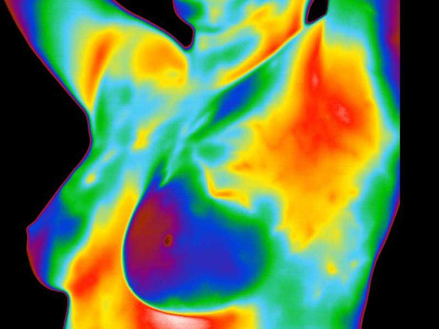
 A breast thermogram
Thermography and the detection of breast cancer
To simply state breast cancer is a mass of genetically malfunctioning cells with excessive uncoordinated growth. Its growth is completely independent from all normal regulatory functions of the host. Breast cancers emerge due to a combination of genetics predispositions, carcinogens, immune responses, hormones, and tissue composition. The breasts are composed of lobes, lobules, ducts, glands, and a high concentration of blood vessels and fat cells. Many of these tissues in the breast have receptors for the hormone estrogen, which makes them a target for the hormone’s influence, particularly the fat cells. Fat cells both produce and breakdown estrogen. The chemical breakdown reaction (aromatization) of estrogen produces carcinogenic (cancer causing) by products. As a result, the carcinogens effect the DNA of nearby cells, which can cause them to mutate into cancers. Research has shown that some women’s breasts are more susceptible than others to the effects of estrogen and its byproducts.
How does the cancer grow?
Once a normal cell begins to mutate (pre-cancerous tissue), its DNA is altered to all for the onset of uncoordinated growth. To sustain the growth of these pre-cancerous (and cancerous) cells, a constant supply of nutrients is needed. In order to maintain this supply, the cells release chemicals into the surrounding area, which keep existing blood vessels open, awaken dormant ones, and create new ones (neoangiogenesis). The rich vascular beds in the breast provide the conditions necessary for the growing tumor’s needs.
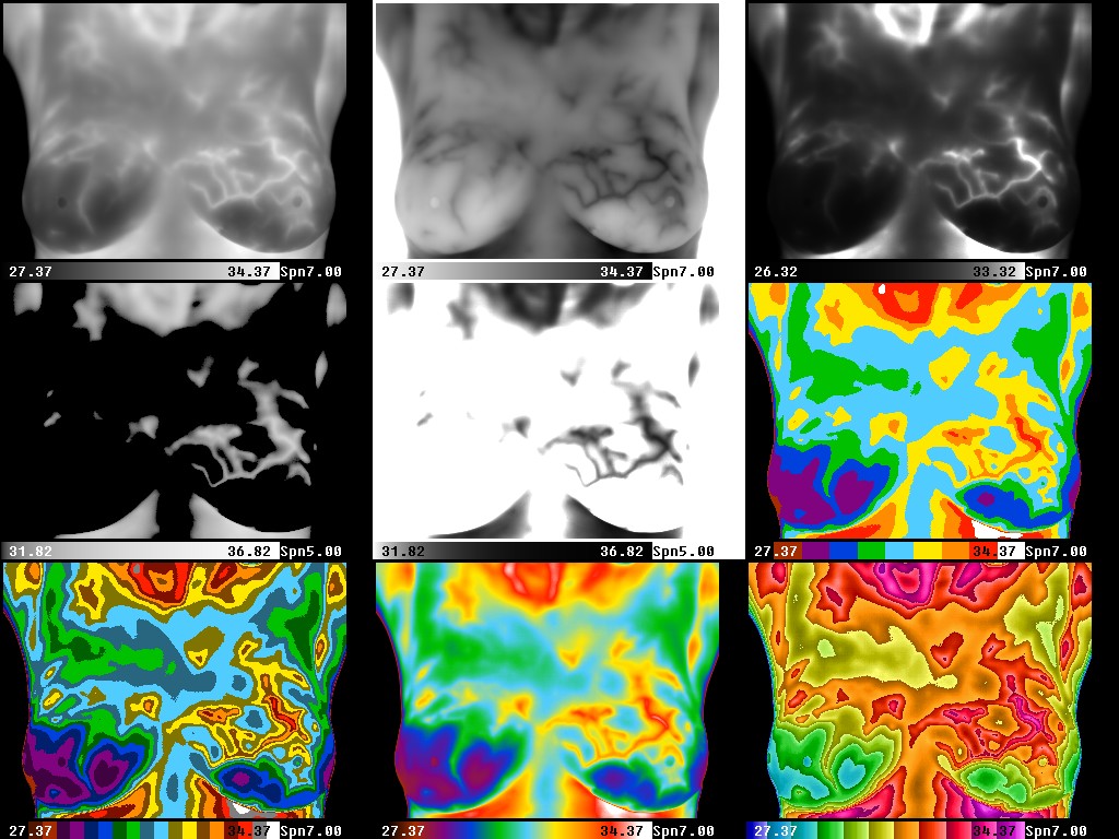
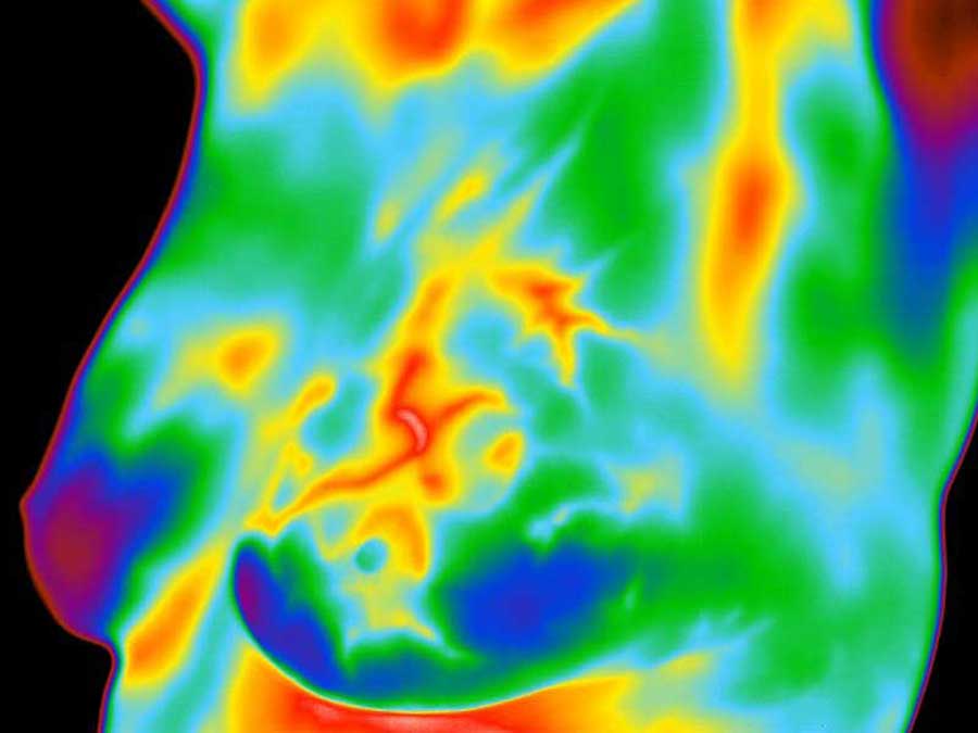 A thermogram showing a blood vessel
How can we detect this growth at Its earliest stages?
Breast cancer is the most common cancer in women, and the risk increases with age (1). Risk is also higher in women whose close relatives have had the disease. Women without children, and those who have had their first child after age 30, also seem to be at higher risk. However, every woman is at risk of developing breast cancer. Current research indicates that 1 in every 8 women in the US will get breast cancer in their lifetime (1).
Studies show an increase in survival rate when breast thermography and structural imaging are used together (2).
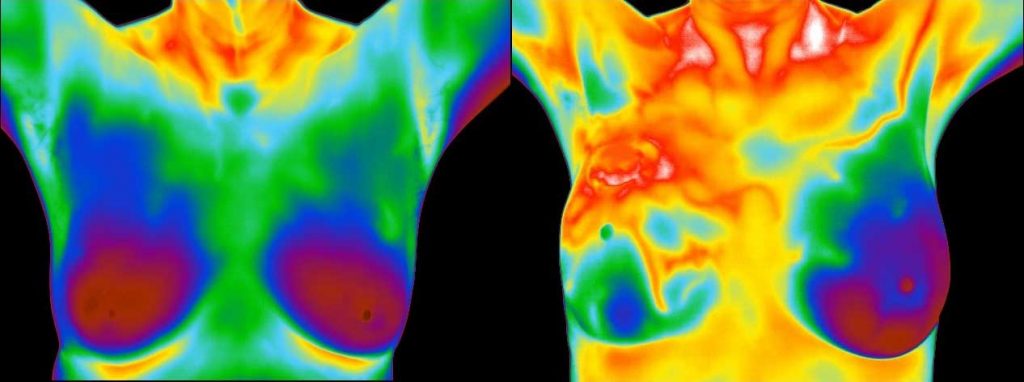 The image on the left is normal, while the image at right shows a severely abnormal breast.
What makes infrared imaging so unique?
While mammography, ultrasound, MRI, and other types of structural imaging rely primarily on finding the physical tumor, thermography is based on detecting the heat produced by increased blood vessel circulation and metabolic changes associated with a tumor’s genesis and growth. By detecting small variations in normal blood vessel activity, infrared imaging may find thermal signs suggesting a pre-cancerous state of the breast or the presences of an early tumor that is not yet large enough to be detected by physical examination, mammography, or other types of structural imaging (2, 3-6).
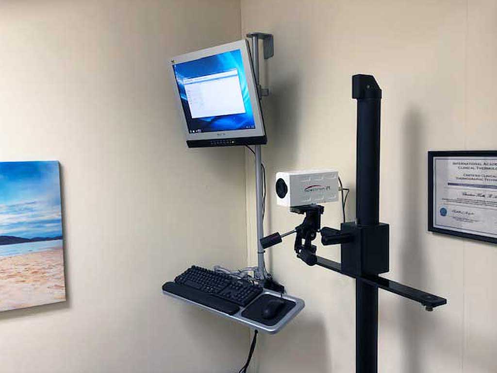 Computerized medical infrared imaging system
Certain types of cancers will not be detected (approximately 20%) by mammography for various reasons (6), but some of these cancers will be discovered by thermography (2, 3-6).
Difficulties in reading mammograms can occur in women who are on hormone replacement, nursing or have fibrocystic, large, dense, or augmented breasts (3,5). These types of breast differences do not cause difficulties in reading infrared images.
How Does Thermography compare to Ultrasound and Mammograms?
Thermography’s ability to detect thermal signs that may suggest a pre-cancerous state of breast, or signs of cancer at very early state, lies in its unique capability of monitoring the temperature variations produced by the earliest changes in tissue physiology (2, 3-6). However, no single imaging technology can offer 100% detection of all early stage cancers. As such, if we are to offer women a method of true early detection, thermography, mammography, ultrasound, and MRI cannot be used alone. These are all adjunctive imaging technologies. Consequently, thermography’s role is in addition to structural imaging and physical examination, not in lieu of. Thermography does not replace any other imaging technology, nor can any other imaging technology replace thermography. In fact, the tests complement each other. Since it has been determined that 1 in 8 women will get breast cancer, we must use every means possible to detect cancers when there is the greatest chance for survival. What we are promoting is a multimodal approach of breast self-exams, physician exams, thermography, and structural imaging together to screen for early detection (2, 4-6). If treated in the earliest stages, cure rates greater than 95% are possible (2,3). With thermography’s expanded role as a risk assessment technology, infrared imaging brings a great deal of good news to women!
How can thermography be used to monitor breast health?
Baseline testing is important to get so you can monitor your health as you age. If A1C (blood sugar average) levels were rising wouldn’t you want to know that you may be at risk for diabetes and to take early measures to prevent getting diabetes, rather than suddenly find out you have diabetes down the road and not even know it was coming? Thermograms of the breast can provide risk markers to give you time to make changes to either prevent the condition or to at least control it. Thermography brings the same risk assessment to the breast health of every woman (7,8). Not only can breast thermography warn that a cancer may be present, but also provide a marker of future risk along with a role in prevention (2,7-11).
The use of thermography is based on the principle that metabolic activity and vascular circulation in both pre-cancerous tissue and the area surrounding a developing breast cancer is almost always higher than in normal breast tissue. Since cancerous tumors have an ever-increasing need for nutrients, an increase in blood circulation is needed. Existing blood vessels are held open, dormant blood vessels are opened and new blood vessels are formed (neoangiogenesis). This process results in and increase in regional surface temperatures of the breast. Thermography uses ultra-sensitive medical infrared cameras and sophisticated computers to detect, analyze, and produce high-resolution images of these temperature variations to find areas of imbalance. Because of thermography’s detection ability, these temperature variations may be among the earliest signs of breast cancer and/or a pre-cancerous state of the breast (2, 3-6).
Current methods used to detect suspicious signs of breast cancer depend primarily on the combination of both physical examination and mammography. While this approach has become the mainstay of early breast cancer detection, more is needed. Since the absolute prevention of breast cancer has not become a reality as of yet, efforts must be directed at detecting breast cancer at its earliest stage. As such, the addition of thermography to the frontline of early breast cancer detection brings a great deal of good news for women.
Who should have this test?
 Mother and daughters
Initial infrared scan by age 20
20-30 years of age — Every 3 years
30 years of age and over — Every year
Every woman should include breast thermography as an addition to monitor her breast health regularly. Everything must be done to provide the earliest detection, in order to prevent women from having to deal with breast cancer.
Breast cancers are particularly aggressive in younger women. Statistics indicate that approximately 15% of all breast cancers occur in women under the age of 45 (12). However, there are no clear guidelines for the use of imaging procedures during these years. With addition of breast thermography, women in this age group have a tool that they can add to their regular breast health check-ups!
References:
- American Cancer Society – Breast Cancer Guidelines and Statistics, 2009-2010
- M. Gautherie, Ph.D.; Thermobiological Assessment of Benign and Malignant Breast Diseases. Am. J. Obstet. Gynecol., 1983; V 147, No. 8: 861-869.
- P. Gamigami, M.D.; Atlas of Mammography: New Early Signs in Breast Cancer. Blackwell Science, 1996.
- J. Keyserlingk, M.D.; Time to Reassess the Value of Infrared Breast Imaging? Oncology News Int., 1997; V 6, No. 9.
- P.Ahlgren, M.D., E. Yu, M.D., J. Keyserlingk, M.D.; Is it Time to Reassess the Value of Infrared Breast Imaging? Primary Care & Cancer (NCI), 1998; V 18, No. 2.
- N. Belliveau, M.D., J. Keyserlingk, M.D. et al ; Infrared Imaging of the Breast: Initial Reappraisal Using High-Resolution Digital Technology in 100 Successive Cases of Stage I and II Breast Cancer. Breast Journal, 1998; V 4, No. 4
- C. Gros, M.D., M. Gautherie, Ph.D.; Breast Thermography and Cancer Risk Prediction. Cancer, 1980; V 45, No. 1: 51-56.
- P. Haehnel, M.D., M. Gautherie, Ph.D. et al; Long-Term Assessment of Breast Cancer Risk by Thermal Imaging. In: Biomedical Thermology, 1980; 279-301.
- Verzini, L., Romani, L., Talia, B., (Radiology department university of Modena (Italy)). Thermographic variations in the breast during the menstrual cycle. Acta Thermographica, 143–149, 1980s.
- Huber, C., Pons, J., Pateau, A., (Gynecology and department of radiology Cretei hospital (Paris) France). Breast fibrocystic disease and thermography. Acta Thermographica, 48–50, 1980s.
- Borten, M., Ransil, B. et al. (Department of obstetrics and gynecology, Beth Israel Hospital, Harvard Medical School). Regional differences in breast surface temperature by liquid crystal thermography. Thermology, 1, 216–220, 1986.
- American Cancer Society – Breast Cancer Guidelines and Statistic.